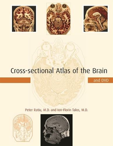

Cross-sectional Atlas of the Brain and DVD
By
Peter Ratiu (Author) Ion-Florin Talos (Author)
Hardback
Available / dispatched within 1 - 4 weeks
Quantity
Description
Cross-sectional Atlas of the Brain provides for the first time a set of high-resolution color cross-sections of the human brain (six times higher than that of the only complete data set available to date), each image accompanied by state-of-the-art MRI and CT scans of the same specimen. The sections were made at an interval of 147 micrometers of frozen tissue, virtually artifact free, with the blood vessels filled at sub-millimeter level. The more than two hundred detailed and fully annotated images in this atlas provide a complete body of reference to the gross anatomy of the brain. The accompanying line drawings of these images provide a roadmap for easy orientation.
The unparalleled resolution of the images also made it possible to derive cross-sections of the same specimen in all standard orientations--sagittal, coronal, and axial--through multi-planar computer-aided reformatting. This feature, which eliminates inter-subject variability, has never before been available in an anatomical atlas and makes the atlas especially useful for identifying and following anatomical structures in each plane. About the Companion DVD(View a sample in PDF format)
While the book itself contains 93 images (44 axial, 28 coronal, and 21 sagittal), the DVD contains the complete series of 1,481 axial images from one anatomic specimen from which the 44 axial images in the book were selected. These images were made at a resolution of 1525x1146 or 147 ?m/pixel with a digital camera. The axial images are accompanied by 1,528 sagittal and 1,146 coronal images that were made by reformatting and reslicing the axial images. By placing these images side-by-side-by-side the DVD allows the user to see a particular region of the brain in all three orientations-axial, sagittal and coronal-simultaneously. These images are further accompanied by radiologic data. The DVD also allows the user to view a synchronized slide show of the images in all three planes. Images on the DVD that also appear in the book are highlighted with a blue background.
Cross-sectional Atlas of the Brain will be an essential reference for neuroscientists and clinicians (neurologists, radiologists, and neurosurgeons). The atlas contains 92 images (44 axial, 28 coronal, and 21 sagittal); the DVD contains 1481 axial images CPSIA choking or other US hazard warning - No California Proposition 65 hazard warning necessary
About the Author
Peter Ratiu, M.D. is Assistant Professor of Radiology at Brigham and Women's Hospital and Harvard Medical School. He is an instructor in Harvard Medical School's course on the Human Nervous System and Behavior. Ion-Florin Talos, M.D. is Instructor in Radiology at Brigham and Women's Hospital and Harvard Medical School. He is an instructor in Harvard Medical School's course on the Human Nervous System and Behavior.
More Details
- Contributor: Peter Ratiu
- Imprint: Harvard University Press
- ISBN13: 9780674019232
- Number of Pages: 240
- Packaged Dimensions: 216x279mm
- Format: Hardback
- Publisher: Harvard University Press
- Release Date: 2006-03-02
- Binding: Hardback
- Biography: Peter Ratiu, M.D. is Assistant Professor of Radiology at Brigham and Women's Hospital and Harvard Medical School. He is an instructor in Harvard Medical School's course on the Human Nervous System and Behavior. Ion-Florin Talos, M.D. is Instructor in Radiology at Brigham and Women's Hospital and Harvard Medical School. He is an instructor in Harvard Medical School's course on the Human Nervous System and Behavior.
Delivery Options
Home Delivery
Store Delivery
Free Returns
We hope you are delighted with everything you buy from us. However, if you are not, we will refund or replace your order up to 30 days after purchase. Terms and exclusions apply; find out more from our Returns and Refunds Policy.